X-ray

First Every X-Ray by Wilhelm Röntgen
|
In Radiology it is noticable how much medicine is bound to other branches of science, specifically physics. Wilhelm Conrad Röntgen who first detected and produced electromagnetic radiation in a wavelength range known as X-rays, was a physicist and a mechanical engineer. That's why "Röntgen rays" is sometimes used to describe X-rays. Today millions and millions of X-ray images are taken for diagnostic and treatment purposes all around the world, from the most advanced centers in the US to remote and far off clinics in underdeveloped countries. Radiology, including X-ray images, revolutionized medicine in a way that no one in the 19th century and before ever imagined; the ability to see inside the body without cutting through. Röntgen was awarded the first ever Nobel Prize in physics in 1901. Do you think a single Nobel Prize is enough to appreciate such a great achievement?
|
Can Pregnant Women Get X-Rays?
|
Have you ever been to a Radiology Department to take an X-ray? If yes, you may have noticed a sign on the wall asking pregnant patients to inform their situation to the staff. What is this for? Is the radiation dangerous for the fetus or the mother? The answer is yes and no. It depends on the age of pregnancy, the amount of radiation and what part of body is going to receive the X-ray. Although it is said that the chance of an X-ray during pregnancy causing harm to the fetus is very small, I believe it is wise to be vigilant. If the staff know the situation, they can adjust the dose of radiation and if the imaging is for some part of the body other than abdomen, they can place a lead apron on the belly to block the passage of radiation to the fetus. Large amounts of radiation in the first two weeks of pregnancy can lead to miscarriage. X-ray, if not necessary, is better to be avoided in the first three months of pregnancy. It may cause growth retardation of the baby or even birth defects. It also may increase the risk of a learning or intellectual disabilities.
|

|
Why Do People Wear Lead Aprons for X-Rays?

|
If X-ray can be potentially dangerous, then how do radiology staff protect themselves from X-ray beams? I think everyone has heard of Lead as an effective material blocking the passage of rays. Why is lead so effective in this matter? Lead is a metal with high molecular density and a large number of electrons. When the radiation wants to pass through lead, its electrons absorb and scatter the energy. That’s why the walls surrounding an X-ray room contain lead. Radiology staff wear aprons having lead in it and you may even see some wearing lead glasses. lead is a very heavy metal. Lead aprons make people look like Captain Phasma in Star Wars! But good news is that there are other, less heavy materials with the same radiation scattering effect of lead: antimony, tungsten, bismuth, and tin also block the rays and are the new generation of radiation protecting materials.
|
X-Rays During Emergencies
|
Do you know what X-ray images are necessary to be taken as soon as possible when a patient with multiple traumas, such as a car accident or fall from a height, is taken to the emergency department? Chest X-ray and a pelvic X-ray are the most necessary imaging and should be taken as soon as the patient's condition permits. The bone and soft tissue shadows seen in these two X-rays help detect life threatening pathologies such as blood or air accumulated in chest (hemothorax and pneumothorax) and pelvic fracture. Early diagnosis of life threatening pathologies through imaging techniques and proper treatment according to those diagnoses help save lives in emergent situations.
|

|
Why Are Multiple Images Needed from the Same Part?

|
Radiology is a branch of medicine; however, radiology, as an imaging technique, needs the sense and finesse of visual art like photography or painting. In an X-ray image, just like photography and painting, you are building a two dimensional image out of a three dimensional reality. This will obscure some details that falls behind other shadows. It is especially true for some complex structures in the body such as the pelvis or the spine. For a long time, this obstacle was a point of concern. One solution to this problem is now to obtain images from different angles so that if a pathology is obscured behind another structure, it can be discovered when you have an image from another angle. That is why so many different angles are needed to view structures such as the pelvis or the spine.
|
Other Technologies have come here to help. Yes, technology gives us thin cross sections of body parts just like slices of bologna sausage. In this way you can have much more details and will be able to detect much more pathologies in the body. MRI, CT Scan, and Ultrasonography are all imaging modalities based on cross section images of the bodies, though only CT Scan uses electromagnetic radiation like X-ray.
Ultrasonography

|
Ultrasonography is another imaging technique to see through the skin and detect the source of disease, or check the wellbeing of an unborn child. This technique uses sound waves to create image. How can this be possible-a conversion of a sound waves into an image?? Again physics comes here to help. The frequency of the sound waves used in ultrasonography is high and out of the range of audibility. The audible range of sound waves are 20 Hz to 20 kHz but ultrasonography uses sound waves with frequencies higher than 20 kHz. The machine sends sound waves into the body using a probe. Different tissues in the body have different reflection properties. The sound waves echo off the tissues depending on this different reflection property which helps radiologists to distinguish between different body organs and pathologies on the produced images.
|
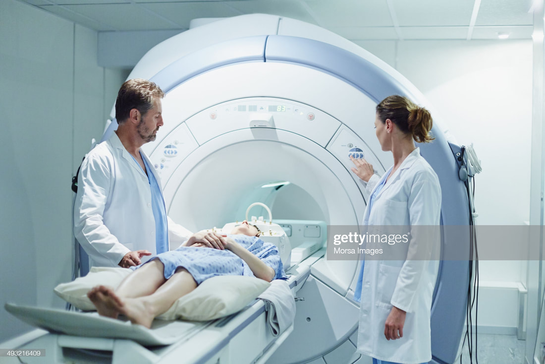
MRI
|
Magnetic Resonance Imaging (MRI) is another imaging modality. Like X-ray, CT scan, and ultrasonography, This technique too, produces images of different body parts. MRI doesn't use electromagnetic waves to produces images but strong magnetic field; just like a huge horseshoe magnet (just kidding)! If you have ever had a sport injury, your doctor may have ordered a knee or ankle MRI for you. The gadget is a huge tunnel that you have to go inside of. The images usually take some time to be taken. There is a lot of noise inside the tunnel, so the staff may give you an earmuff to block the noise. Once I went through this and the staff played some music for me through the earmuff! If you are lucky the earmuff may play some nice music for you too (In case an injury happens). Despite all these measures, some cannot tolerate that situation and may panic due to claustrophobia.
|
|
But How Does MRI Actually Work?
It is pure physics and really not easy to explain. Hydrogen atoms come to help in this matter. You know there are abundant hydrogen molecules especially in water and fat, and every organ in the body has different concentrations of water and fat. That is why MRI images are essentially the map of water and fat distribution in the body. When the body goes inside the strong magnetic field of MRI machines, all hydrogen molecules in the body align themselves in the magnetic field direction. Pulses of radiofrequency current are then sent to the body and cause the hydrogen protons go out of equilibrium. The radio waves are then stopped and the hydrogen molecules realign again with the magnetic field. The speed of this realignment of protons is different depending on the environment and the chemical characteristics of the molecules. This difference is printed as an MRI image showing different body organs.
How Do Doctors Remove Foreign Items Out of the Body?
Phew! Now lets talk about a more simpler topic. Sometimes a foreign object enters the body through the skin and causes pain and problems. How can a doctor find that intruding object and take it out. Here again imaging modalities help. Can we use an X-ray to detect a foreign body? It depends on that object and if it is radio dense or radiolucent, simply meaning that if the object can be detected by an X-ray or not. Metal-containing pieces are easily seen in X-ray, such as a needle. Glass particles are sometimes seen and sometimes not. How about a splinter of wood? I have to say unfortunately you cannot see a chip of wood with an X-ray. So what should you do? You Guessed it! Again it is MRI that does the job. With the same mechanism explained in the previous topic, pieces other than body parts can be easily distinguished on MRI and removed if needed.
